This blog post was originally published at e-con Systems’ website. It is reprinted here with the permission of e-con Systems.
Microscopic-based diagnostic devices are critical, but they come with many imaging challenges. Explore how to overcome these challenges effectively and their popular use cases. Also learn about a 5MP high-sensitive camera by e-con Systems – tailor-made for microscopic-based diagnostic devices.
Microscopic-based diagnostic devices have revolutionized medical diagnosis, enabling detailed examination of cellular and tissue samples. Cameras integrated into these devices are critical in visualization, documentation, and analysis. However, various imaging challenges exist in different medical segments.
In this blog, let’s explore the major use cases, their challenges, and the types of cameras required to address them effectively. You’ll also learn about See3CAM_50CUG – a 5MP high-sensitive camera by e-con Systems that can amplify the performance of microscopic-based diagnostic devices.
Role of cameras in microscopic-based diagnostic devices
Cameras have emerged as indispensable tools in advancing microscopic-based medical diagnosis. With their high-resolution sensors and advanced imaging capabilities, cameras enable healthcare professionals to capture and analyze detailed microscopic images with unprecedented clarity. These images serve as valuable visual evidence, aiding in the accurate identification and characterization of cellular structures, tissue samples, and pathogens.
Furthermore, real-time visualization and image analysis facilitated by cameras empower medical practitioners to make informed decisions, improve diagnostic accuracy, and enhance patient care.
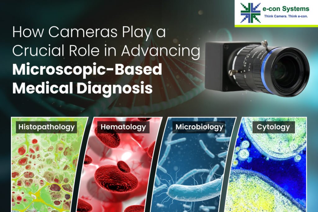
Major uses cases of microscopic-based diagnostic devices
Histopathology
Histopathology is a field that plays a critical role in diagnosing diseases and understanding tissue abnormalities. It involves the examination of tissue samples under a microscope to identify and analyze the changes occurring at a cellular level. By studying the microscopic features of tissues, histopathologists can provide valuable insights into the nature and progression of diseases.
However, histopathology imaging comes with its own set of challenges. The good news is that there are camera solutions to overcome them! Let’s take a look:
| Challenges | Camera Solutions |
| Unable to capture images from large tissue sections | High-resolution cameras with large sensors |
| Unable to achieve uniform illumination and depth of field | Cameras with adjustable depth of field capabilities |
Hematology
Hematology is a branch of medical science that focuses on studying blood and blood disorders. It involves examining and analyzing blood samples to diagnose and monitor various conditions related to blood cells, such as anemia, leukemia, and clotting disorders. In hematology imaging, specific challenges need to be addressed to ensure accurate identification and differentiation of blood cell types. Here is the list of challenges and their corresponding solutions:
| Challenges | Camera Solutions |
| Inaccurate identification and differentiation of blood cell types | Cameras with excellent color reproduction capabilities |
| Lack of precise and high-speed imaging for dynamic blood cells | Cameras with fast frame rates and superior light sensitive image sensors |
Microbiology
Microbiology is a scientific discipline that focuses on studying microorganisms, including bacteria, viruses, fungi, and other microscopic organisms. It plays a crucial role in various fields, such as medicine, agriculture, environmental science, and biotechnology. Microbiology imaging involves visualizing and analyzing microorganisms to understand their structure, behavior, and interactions. However, certain imaging challenges must be addressed to capture clear images of microorganisms, often surrounded by complex backgrounds. Here’s how you can get it done.
| Challenges | Camera Solutions |
| Unable to capture clear images of microorganisms surrounded by complex backgrounds | Cameras with high-resolution capabilities and good dynamic range |
| Inability to enable rapid image acquisition for real-time analysis | Cameras with an accurate high frame rate |
Cytology
Cytology is a branch of biology that focuses on the study of individual cells to understand their structure, function, and behavior. In the medical field, cytology plays a crucial role in diagnosing diseases, particularly cancer, by examining cells for abnormalities and identifying malignant or precancerous conditions. Now, let’s explore their challenges and related camera solutions.
| Challenges | Camera Solutions |
| Lack of visualization and analysis of individual cells within complex samples | High-resolution cameras with low-light sensitivity and excellent signal-to-noise ratio |
| Unable to maintain cell viability and minimize background interference | Cameras with manual exposure control |
Urine sediment analysis
Urine sediment analysis is a diagnostic procedure involving the microscopic examination of cellular elements and structures in urine samples. It is an essential component of urinalysis that helps assess kidney function, detects urinary tract infections, and identifies various renal disorders. During urine sediment analysis, specific challenges need to be addressed to ensure accurate identification and enumeration of cells and structures.
| Challenges | Camera Solutions |
| Inaccurate identification and enumeration of cells and structures | Large pixel size cameras with enhanced contrast and sharpness |
| Unable to visualize small and transparent cellular elements | Cameras with high-resolution imaging and NIR capabilities |
Peripheral blood smear
Peripheral blood smear analysis is a diagnostic technique that examines a thin layer of blood cells spread on a glass slide. It is a valuable tool in hematological investigations and allows for evaluating various blood cell types, their morphology, and any abnormalities present. Here’s a summary of the camera solutions for the challenges faced in peripheral blood smear imaging:
| Challenges | Camera Solutions |
| Unable to capture high-quality images of blood cells with varying morphologies and sizes | High-resolution cameras with excellent color fidelity |
| Unable to capture rare cell types | Cameras with options to select the region of interest (ROI) |
See3CAM_50CUG – a best-fit camera solution by e-con Systems
e-con Systems, a pioneer in the embedded vision space since 2003, offers a wide range of cameras to build cutting-edge medical devices, including diagnostic microscopes. The See3CAM_50CUG proves to be the ideal camera choice to address the imaging challenges of histopathology, hematology, microbiology, cytology, urine sediment analysis, and peripheral blood smears.
This 5MP high-sensitive color camera leverages the Sony Pregius IMX264 CMOS sensor for exceptional image quality and accuracy and comes with relevant features such as:
- High-resolution imaging
- Adjustable depth of field
- Excellent color reproduction
- Advanced image processing capabilities
Learn more:
Check out the Camera Selector page to look at our full portfolio.
To get started with our advanced camera solutions, visit our website or write to [email protected].
Balaji S
Product Manager, e-con Systems